 Breaking news
 Europe
 Bulgaria
A new study by Brown University researchers shows that gold nanoparticles - microscopic pieces of gold thousands of times thinner than a human hair - could one day be used to restore vision in people with macular degeneration and other retinal disorders.
The research was published in the journal ACS Nano and supported by the National Institutes of Health. In it, the research team demonstrated that nanoparticles injected into the retina can successfully stimulate the visual system and restore vision in mice with retinal disorders. The findings suggest that a new type of vision prosthesis system, in which nanoparticles used in combination with a small laser device embedded in a pair of glasses, could one day help people with retinal disorders see again.
"This is a new type of retinal prosthesis that has the potential to restore lost vision in retinal degeneration without the need for complex surgery or genetic modification. We think this technique could change paradigms for treating retinal degenerative conditions," said Jiarui Ni, a postdoctoral fellow at the National Institutes of Health who led the research while completing his doctorate at Brown.
Ni performed the research while working in the lab of Zhonghuang Lee, an associate professor in Brown's School of Engineering and a faculty member at Brown's Carney Institute for Brain Sciences, who led the work and was the study's senior author.
Retinal diseases such as macular degeneration and retinitis pigmentosa affect millions of people worldwide. These conditions damage light-sensitive cells in the retina called photoreceptors - the "rods" and "cones" that convert light into tiny electrical impulses. These impulses stimulate other types of cells further up the visual chain, called bipolar and ganglion cells, which process the signals from the photoreceptors and send them to the brain.
This new approach uses nanoparticles injected directly into the retina to bypass damaged photoreceptors. When infrared light is focused on the nanoparticles, they generate a small amount of heat that activates bipolar and ganglion cells in the same way as photoreceptor impulses. Because diseases such as macular degeneration primarily affect photoreceptors, while bipolar and ganglion cells remain intact, the strategy has the potential to restore lost vision.
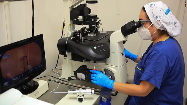
In this new study, the research team tested the nanoparticle approach in the retinas of mice and in live mice with retinal disorders. After injecting a liquid solution of nanoparticles, the researchers used patterned laser light in the near-infrared to project shapes onto the retina. Using a calcium signal to detect cell activity, the team confirmed that the nanoparticles excited bipolar and ganglion cells in patterns consistent with the shapes projected by the laser.
The experiments showed that neither the nanoparticle solution nor the laser stimulation caused detectable adverse side effects, as indicated by metabolic markers of inflammation and toxicity. Using probes, the researchers confirmed that laser stimulation of the nanoparticles induced increased activity in the visual cortex of the mice. This was an indication that previously absent visual signals were being transmitted to and processed by the brain. According to the researchers, this is a sign that vision has been at least partially restored, which bodes well for the eventual application of similar technology in humans.
The researchers envision a system that would combine the nanoparticles with a laser system mounted in a pair of glasses. Cameras in the goggles would collect image data from the outside world and use it to control the modelling of an infrared laser. The laser pulses will then stimulate the nanoparticles in people's retinas, allowing them to see.
The approach is similar to one that was approved by the Food and Drug Administration for human use several years ago. The older approach combined a camera system with a small electrode array that was surgically implanted in the eye. According to Nee, the nanoparticle approach has several key advantages.
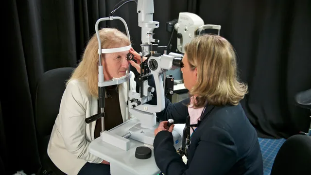
First, it is much less invasive. Unlike surgery, "intravitreal injection is one of the simplest procedures in ophthalmology," Nee said.
The approach is similar to that which was approved by the Food and Drug Administration for human use several years ago. The older approach combined a camera system with a small electrode array that was surgically implanted in the eye. According to Nee, the nanoparticle approach has several key advantages.
For starters, it is much less invasive. Unlike surgery, "intravitreal injection is one of the simplest procedures in ophthalmology," Nee said.
There are also functional advantages. The resolution of the previous approach was limited by the size of the electrode array - about 60 square pixels. Because the nanoparticle solution covers the entire retina, the new approach could potentially cover a person's entire field of view. And because the nanoparticles respond to near-infrared light rather than visible light, the system doesn't necessarily interfere with the residual vision a person can retain.
More work needs to be done before the approach can be tested in a clinical setting, according to Nee, but these early studies suggest it is possible.
"We have shown that nanoparticles can remain in the retina for months without serious toxicity. And we showed that they can successfully stimulate the visual system. This is very encouraging for future applications," said the scientist. | BGNES